Search This Blog

Tuesday, November 16, 2010

Pulmonary renal syndrome

In rheumatology morning report today we discussed an older woman presenting with hemoptysis and acute kidney injury with a Cr in the 700s. This lead to an interesting discussion of the many systemic disorders that can present with renal and pulmonary manifestations.

Pulmonary renal syndrome is essentially the simultaneous occurrence of both glomerulonephritis and diffuse alveolar hemorrhage. This usually manifests as a rapidly rising Cr, hematuria, and shortness of breath/cough/hemoptysis/desaturation. However, we must also remember that other conditions may cause acute kidney injury and pulmonary manifestations as well (ie acute kidney injury from prerenal failure or ATN complicating pneumonia, ARDS/AKI from sepsis/trauma, malaria, rapidly progressing AKI in CHF, etc). What really is needed to differentiate these entitities from true pulmonary renal syndrome is the presence of alveolar hemorrhage on bronchoscopy and the presence of glomerulonephritis on biopsy or urine microscopy (hematuria, red cell casts).

The causes of pulmonary renal syndrome include:

Connective Tissue Disorders

Polymyositis or dermatomyositis

Progressive systemic sclerosis

RA

SLE

Anti-glomerular basement membrane disease

Goodpasture's syndrome

Systemic Vasculitidies

Behçet's syndrome

Churg-Strauss syndrome

Cryoglobulinemia

Henoch-Schönlein purpura

Microscopic polyarteritis

Wegener's granulomatosis

Other

Drug Reactions

We discussed that the initial workup for these patients should include CBC + diff (for cytopenias), smear (for hemolysis), Cr, BUN, Lytes, Ca, Mg, PO4, albumin, LFTs, INR, PTT, CXR, EKG, and urinalysis and microscopy.

Further serology should include:

ANA, dsDNA if ANA positive (for SLE)

ANCA (c-ANCA (anti-PR3) positive in Wegener's, p-ANCA (Anti-MPO) in microscopic polyangiits, P>C-ANCA in Churg-Strauss. (Note: immunofluorescence is sensitive but less specific while ELISA is specific and sensitive. Know which one your lab does.)

Anti-GBM for Goodpasture's

Definitive Diagnosis: biopsy of lung/kidneys.

It is often necessary to make quick but definitive diagnoses as the consequences of not treating are dire, but the side effects of many of our immunosuppressive agents are not insignificant.

Please see a post from July on an interesting case of levimasole-induced vasculitis.

See here for an interesting review of ANCA vasculitidies from 2009.
Tuesday, November 9, 2010

Varicella & Pregnancy


Last week we discussed a case of an immunosuppressed woman with herpes zoster infection of the face, and the concerns that go along with that (what to do with her immunosuppressive medications? Ophthalmalogic concerns? Differential diagnosis). Today, we discussed a woman visiting from sub-Saharan African with a diffuse vesicular rash, ultimately thought to be disseminated varicella with varicella pneumonia. A surprise to everyone - someone wisely ordered a BHCG and she was, in fact, pregnant.

The CDC "Pink Book" has a wealth of information on many vaccine preventable illnesses - easily found by googling "CDC Pink Book". Looking under varicella, there is access to pictures, literature, vaccine recommendations, and more. Here is the link.

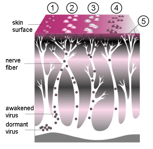
Varicella zoster virus (VZV) is a member of the herpesviridae family responsible for both acute infection ("chickenpox") and recurrent infection (herpes zoster, or "shingles"). After primary infection, VZV persists in sensory nerve ganglia and can result in recurrent infection, especially in immunocompromised hosts. It is transmitted through the respiratory tract (airborne & contact precautions in most hospitals!) with an incubation period of 14-16 days, although longer in immunocompromised hosts. Primary infection, as in our patient, is usually preceded by a 1-2 day malaise, followed by a generalized, pruritic rash that progresses from macules, to papules, to vesicular lesions, then crusts. Lesions of all ages can be found. Recurrent infection is more common in those with immunosuppression, advanced age, intrauterine VZV exposure, and VZV infection at less than 18 months. Zoster can become disseminated and involve the skin, CNS, lungs, and liver. It typically involves the fifth cranial nerve (as in our patient last week) or the trunk.

Complications can be severe, and can include:
  • Secondary bacterial infection of skin lesions
  • Viral or secondary bacterial pneumonia
  • Meningitis or encephalitis (especially affecting cerebellum, leading to ataxia)
  • Reye syndrome
  • Myocarditis, GN, Transverse Myelitis, TTP, adrenal insufficiency
  • Congenital Varicella Syndrome (CVS)
Congenital Varicella Syndrome is a risk if infection occurs in the first 20 weeks of gestation. Risk is actually pretty low (2%). It is manifest by:

  • Low birth weight
  • Hypoplasia of an extremity
  • Skin scarring
  • Localized muscular atrophy
  • Encephalitis, cortical atrophy, chorioretinitis, and microcephaly

CVS is distinct from the risks if mum develops VZV from 5 days before to 2 days after delivery, which can result in overwhelming neonatal infection and 30% fatality rates. This is thought to be the result of fetal exposure without passive maternal antibodies.

We consider VZIg in certain populations, including:

  • Immunocompromised patients
  • Neonates whose mothers have signs and symptoms of varicella around the time of delivery
  • Preterm infants born at or greater than 28 weeks gestation who are exposed during the neonatal period to non-immune mums
  • Preterm infants born at less than 28 weeks' gestation or who weigh 1,000g or less at birth and were exposed during the neonatal period, regardless of maternal history of varicella disease or vaccination
  • Pregnant women within 96 h of exposure
A few final pearls:
  • All pregnant women should receive treatment (ie with acyclovir). See here for an article on management of VZV in pregnancy.
  • Don't forget to consider other possibilities: Measles? Coxsackie? Impetigo? See here for some pictures and a summary table.
  • Lastly, we must remember that geography plays a large role in varicella immunity. Many immigrant and refugee populations come from tropical and subtropical climates, where varicella immunity is much LOWER than in temperate climates like ours. Thus, a pregnant mum hailing from Zambia is a lot less likely than one born in Toronto to be immune to VZV. Given the risks, we should perhaps be engaging in routine antenatal screening of women from tropical/subtropical zones and providing vaccination, as well as insuring that they have access to timely and appropriate antenatal/pre/postnatal care. There has been some study on this issue, and here's an example.

Thursday, November 4, 2010

Low Back Pain


Today we discussed a common presentation but difficult dilemma: chronic low back pain, with new neurological symptoms. When do you know to investigate further? How do you manage complex pain? What are the red flags? What is an anatomical approach to neurological manifestations? We discussed many of these issues today. For the internist, it is often important to be able to differentiate inflammatory from mechanical back pain.

It's worthwhile discussing the differential diagnosis of back pain, which includes:
Mechanical
  • MSK injury
  • Degenerative discs & facet joints
  • Anatomic anomalies (scoliosis, spondylolisthesis)
Neurological
  • Disk herniation, with irritation of adjacent nerve roots
  • Spinal stenosis
Non Mechanical Spinal
  • Malignancy (primary or secondary) with or without cord compression
  • Inflammatory: Ankylosing spondylitis (& other seronegative spondyloarthropathies), RA
  • Infectious: osteomyelitis, epidural abscess
Visceral radiation (pelvic, kidney, GI tract, aorta)

Remember that there are several key elements to a thorough back examination:

Inspection: anteriorly (looking for alignment), posteriorly (looking for asymmetry, deformity, rashes, etc), and laterally (for kyphosis, lordosis). Also inspect their gait.

Range of motion: Assess flexion, extension, rotation, and lateral bend while standing, looking for limitation & pain. From the seated position, test rotation of the spine at the thoracolumbar region - limitation is an early indicator of inflammatory back pain.

Palpation: of spinous processes and paraspinal muscles for pain or spasm.

Special Tests: compression test (of SI joints) with patient on side, FABER test, Gaenslen's test, Book test with patient supine (pressure over both trochanters.
Other special tests to differentiate between mechanical and inflammatory back pain include the straight leg raise (positive in mechanical), femoral stretch test, the occiput to wall test, the Modified Schober, and the

A full neurological examination should accompany this exam, including a DRE to assess for sphincter tone if cord compression is suspected.

See here for an excellent BMJ review.

Wednesday, November 3, 2010

Mental Health, Incarceration, and COPD


Thanks to Dr. Sargeant for an interesting morning report today, allowing us to explore some of the more complex issues affecting our patients with mental health concerns, including their disproportionately high representation in the prison system. The causality of this relationship is complex, and often influenced by confounders like addiction, but ultimately we have to look at this as a matter of public policy and how Canada (and other nations around the world) have chosen to allocate resources. I found an interesting resource document (here) from the American Psychiatric Association that discusses some of the broader factors and implications of the relationship between mental illness and the criminal justice system. An article on the role played by addiction can be found here.

We also discussed the clinical examination for airway obstruction, focusing on Dr. Sharon Straus' practical approach outlined here. Essentially, the combination of (all three) 1) self reported COPD, 2) Forced expiratory time >9 seconds, and 3) wheeze on auscultation had a positive likelihood ration (see here for definition) of 59, essentially ruling in COPD. Patients with none of the three findings had a negative LR of 0.3, effectively ruling it out.

Thursday, October 21, 2010

HIV, Splenomegaly & Lymphoma


Today we discussed the case of an HIV positive man presenting with "L flank pain" found to have anemia, splenomegaly, and a left renal vein thrombosis.

There are many learning points from this case:

The differential diagnosis for splenomegaly:
  • Reticuloendothelial hyperplasia (chronic hemolysis, sickle cell, thal major)
  • Infection-related hyperplasia (including HIV, EBC, CMR, TB, malaria, MAI)
  • Autoimmune hyperplasia (SLE, RA - known as Felty's syndrome when neutropenia present)
  • Malignancies (myeloproliferative, leukemia, lymphoma, myeloma, amyloid)
  • Granulatomous disease (Sarcoid, TB)
  • Congestion (Cirrhosis, CHF, portal or splenic vein thrombosis, schistosomiasis)
  • Infiltration (storage diseases)
The rational clinical exam for splenomegaly (see here) that suggests that palpation and percussion are most helpful if your pretest probability is >10%

The clear association between HIV and lymphoma, most commonly high-grade B-cell non-Hodgkin's lymphoma. However, increasingly we are seeing more and more Hodgkin's lymphomas in HIV+ patients as well. See here for more details.

Hyponatremia and psoas abscess


Last week we discussed a patient with chronic liver disease secondary to alcohol presenting with gram-negative bacteremia, hyponatremia, and a psoas abscess. We focused mostly on the diagnosis and management of hyponatremia.

Hyponatremia: there are a few questions you must ask yourself:

1. Is it real?
  • Think of other osmoles - glucose, mannitol, paraproteins
2. Is the urine reponse appropriate - ie what is the Urine Osmolality?
  • If low, the urine is appropriately dilute
  • If > 100, it is inappropriately concentrated - there must be ADH on board
3. What is the patients volume status?
  • Determine if hypo-, iso-, or hypervolemic
  • Physical exam is helpful in the extremes, but interobserver reliability is low.
  • In the absence of diuretics, Urine Na can be helful - less than 20 suggests the kidneys are avidly reabsorbing Na, so the patient is either hypovolemic or has low effective circulating volume but hypervolemic. The physical exam is helpful in differentiating these states.
  • If the patient is on a diuretic, the fractional excretion of urea can be used (value >35% is euvolemic). Defined as Fractional Excretion of Urea (FEUrea) = (SerumCr * UUrea ) / (SerumUrea x UCr) %.
Once you have answered these three questions, you can accurately classify their hyponatremia as hypovolemic (renal or extra-renal losses, the urine Na can help determine), euvolemic (SIADH based on your urine Osm, adrenal insufficiency, hypothyroidism, or primary polydipsia/low solute diet if the urine is appropriately dilute), or hypervolemic (ie liver, cardiac, or renal failure).

In managing hyponatremia, it is essential to avoid overly rapid correction which can cause osmotic demyelination syndrome. Treatment modality will depend on the patients volume status. There are very few indications for hypertonic saline - essentially only if the patient is seizing and you think it's related to their sodium. Remember to monitor the sodium level frequently as well as the urine output - once you have volume repleted the patient, the stimulus for ADH gets 'shut off', resulting in sudden outpouring of dilute urine which can cause a sudden (too quick!) rise in your serum sodium.

Here is a brief review of hyponatremia.

If you are interested in learning more about psoas absecesses, take a look at this review.

Wednesday, October 6, 2010

Fever of Unknown Origin


After a month of junior attending, the blog is back! On Tuesday we discussed an interesting case of fever of unknown origin (FUO). FUO is a clinical entity that is defined as a T > 38.3 on several occasions for >3 weeks, the etiology of which remains unclear after 1 week of diagnosis.

A home-grown review article (see here) highlights the importance of approaching these patients methodically. Of note, this algorithm does not apply to patients who are HIV+ or have a known malignancy, or to children. Up to 20% of patients with FUO remain undiagnosed. Among those in whom a diagnosis is found, 25-30% are found to have infectious causes, 20-30% are found to have inflammatory causes, and 15-20% have malignant causes. The remainder belong to the "other" category - see below.

Infectious Causes of FUO
Bacterial
Endocarditis (apply Duke criteria, think of HACEK organisms and Q fever)
Abscesses (especially intra-abdominal - consider CT)
Dental abscesses, sinusitis
Osteomyelitis
Mycobacteria (TB)
Spirochetes (Syphilis - get VDRL, Lyme disease)

Viral
HIV, HBV, HCV, EBV, CMV

Fungal
Aspergillus, histoplasmosis, coccidiodomycosis, blastomycosis, disseminated candida

Parasites
Malaria, schistosomiasis, Chagas

Inflammatory Causes of FUO
SLE, RA, PMR, Sjogren's
Vasculitis (GCA, WG, PAN)
Behcet's Disease
Sarcoid, IBD, Still's Disease

Malignant Causes of FUO
Leukemia, Lymphoma
Renal Cell CA
Hepatocellular CA
Colorectal CA

Other Causes of FUO
Drug fever
DVT/PE
Subacute thyroiditis
Familial mediterranean fever
Factitious
Disorder of hemostasis

Key principles and approach (see article link above for a great algorithm):
1. Take a thorough Hx and P/E to point you to a focus: thorough ROS, travel/medication/sexual/occupational history, infectious risk factors
2. Stop all non-essential medications and fight the urge to treat empirically
3. Basic investigations: CBC + diff + film, Cr, LFTs, anti-HCV, HBsAg, anti-HBs, Blood cultures x3, UA and urine culture, HIV, CMV/EBV serology may be helpful, ANA, RF, LDH, CXR
4. Abdo CT to rule out abscess and look for tumour, nodes +/- Technitium-based nuclear scan to identify a focus
5. Apply duke criteria and obtain echo, blood cultures
6. Leg dopplers to rule out DVT
7. If older than 50, get temporal artery biopsy to rule out GCA
8. Liver bx can be helpful if LFTs abnormal

Wednesday, September 1, 2010

Polyuria and hyperosmolar hyperglycemic state


Today we discussed an 80 year old many with polyuria and weakness, found to have a hyperglycemic hyperosmolar state, or HHS (previously HONK).

We discussed the differential diagnosis of polyuria:
If urine osmolarity = (# of particles/urine volume), urine vol must = # of particles/urine osm.
Thus, polyuria is a problem of either too many particles or too low urine osmolarity.

Too many particles:
Glucose in DM
NaCl in people on diuretics or post-ATN or with a salt-wasting nephropathy
Urea in people receiving tube feeds

Too low osmolarity:
Primary polydipsia
Diabetes Insipidus

We also discussed the various causes of diabetes insipidus. See here for a review of DI that highlights these causes nicely.

Lastly we discussed HHS and its management. Patients with HHS are often profoundly volume deplete (up to 10L), with sugars much higher than in DKA. Although some advocate for treatment without insulin, in most situations IV insulin is required or helpful. I can't summarize it better than the great algorithm in this article - see here.

Tuesday, August 24, 2010

Systemic Lupus Erythomatosus


Today we had the chance to briefly discuss the new diagnosis of SLE in a South-East Asian male presenting with fever, chills, pleural effusions, and a drug-induced maculopapular rash. He was found to have pancytopenia (including both neutropenia and lymphopenia), an elevated creatinine, pancolitis on CT, pleural and pericardial effusions.

The ACR criteria for lupus suggest that patients must have 4 of the 11 criteria, although not necessarily concurrently. This lack of "simultaneous manifestations", unlike in our patient, can make earlier SLE much harder to diagnose without a high index of suspicion. This is important because the later the diagnosis, the more chance for irreversible damage, such as worsening lupus nephritis.

Here is an article on sex disparities in SLE. Interestingly, 4-22% of SLE occurs in males. Case control/cohort studies suggest that men experience more renal disease with SLE, and also more hematological manifestations. Serositis may also be more common in men. However, these results are conflicted and may be more complex in terms of race interactions and other factors than initially believed. Men may have a poorer long-term prognosis than women suggested by higher disease activity scores (SLICC score).

SLE, perhaps more so than any other rheumatological condition, is manifest by gross disparities in outcomes among patients of varying socioeconomic class and race/ethnicity. This has been extensively studied. Many factors play a role, inlcuding fixed factors, such as age, sex, race, and geography, and modifiable factors such as access to healthcare, insurance, social support. It is important to remember that these disparities exist in a sociopolitical context as well. Many texts classify socioeconomic status as a fixed factor, but I would argue that it is potentially modifiable with more adequate social assistance, and policies that recognize and address the social determinants of health. Here is a review of the disparities in SLE.

Wednesday, August 18, 2010

Non-typhoidal salmonella bacteremia and HIV


Morning report today focused on an HIV+/HBV coinfected man with a CD4 count of 7, presenting with diarrhea, cachexia, and cirrhosis following travel, ultimately diagnosed with Salmonella enteriditis bacteremia.

Salmonella infections can be classified as typhoid fever, or those caused by Salmonella typhi or paratyphi, and other non-typhoidal salmonella (NTS) infections. NTS clinical syndromes can take 4 forms:
1) diarrheal disease (colitis)
2) invasive bacteremia
3) focal or suppurative infection
4) asymptomatic carriage in stool.

HIV+ patients are at much higher risk for invasive NTS bacteremia when compared to HIV negative populations, and are much more likely to have recurrence of disease. Recurrent often occurs with the same strain and is thought to be related to reticuloendothelial persistence. The presentation can be quite non-specific and may not include any diarrhea at all.

See here for a brief review article on salmonella in immunocompromised hosts.

Treatment is typically with fluoroquinolones. Plasmid-mediated resistance does exist, but in relatively few isolates in the US - our patient was successfully treated with cipro as well. See here for a study about this if bugs and drugs are your thing!

Lots more to discuss: differential diagnosis of esophageal ulcers in and HIV+ patient, differential diagnosis of diarrhea in HIV+ patients, and HBV/HIV coinfection, but I'll have to save those for future blogs! Thanks to Dr. Vellend for being our guest.

Tuesday, August 17, 2010

On being a good physician


Today with Dr. Kitchens we had the special privilege of reflecting on our memorable cases and our most recent reads. It was great to hear that we all indulge our passions and interests outside of medicine, whether it is through stories of harpooning and sinking ships or reading to your loved ones.

The medicine teams right now are reading:

My name is red by Orhan Pamuk
The Kiterunner by Khaled Hosseini
Complications by Atul Gawande
A Spot of Bother by Mark Haddon
Never Let me Go by Kazuo Ishiguro
Consumption by Kevin Patterson
Omar Khayyam's Rubaiyyat
Among others.

I'm glad we had the chance today to reflect on what makes us good physicians, and not just good doctors. Thanks Dr. Kitchens!

I'll leave you with some of the poetry we heard about today (Omar Khayyam):

Here with a Loaf of Bread beneath the Bough,
A Flask of Wine, a Book of Verse, and Thou
Beside me singing in the Wilderness
And Wilderness is Paradise now.

Monday, August 16, 2010

Palliative care


Today we had an introduction into palliative care by Dr. LaDelfa.

Often, in medicine, we are called to support individuals through extremely ill health - and often we focus on trying to stall, postpone, or even prevent death. As physicians, though, we can sometimes play an even more important, kinder, and helpful role - that of facilitating a dignified and comfortable death.

Dr. LaDelfa highlighted some of the key elements of end of life care.

1. Provide effective symptom management
2. Help restore a patient's sense of control over their own life
3. Provide patients with the opportunity to deal with their existential concerns (psychological, religious)
4. Help patients feel relieved from their physical, financial, and emotional burdens
5. Provide patients with (or facilitate) time to engage in discussions with their families and friends to affirm their lives - that they had meaning, that they experiences successes, experienced love.

Please see here for a wonderful essay written by Atul Gawande in the New Yorker on "Letting Go" - on how liberating, for patients and providers alike, the experiences of death and dying can be when we focus on quality of life rather than quantity. I feel as though this should be required reading for all medical practitioners!

Friday, August 13, 2010

Cardiac tamponade


Thanks to Dr. Parker for taking us through some really interesting echocardiographic findings in a patient with cardiac tamponade.

Tamponade occurs when the intrapericardial pressure rises to the point where it compromises venous filling of the right atrium, which can actually be seen to collapse on echocardiogram. Impaired blood flow to the right side of the heart means less blood to go to the left side, exacerbated by the RV's attempt to expand in a compressed space which leads to interventricular bulging into the LV. This decreased LV filling results in the clinical findings of hypotension and pulsus paradoxus. Acute tamponade is immediately life-threatening and will require pericardiocentesis - get an echo and a cardiology consult!!!

See here for a recent review of pericardial diseases.

Thursday, August 12, 2010

Hypertension


On thursday we discussed an interesting patient with possibly autonomic dysreflexia, possibly due to a prior cord injury. This led to an interesting discussion of hypertensive urgency and its management.

Understandably, on internal medicine we often focus on hypertensive urgencies (typically >180/110 with no impending progressive end-organ damage) and emergencies (uncontrolled BP with acute progressive end-organ damage) because they are what we manage as an inpatient. Here is a recent review article on the management of these entities.

However, in the ambulatory setting we are often asked to initiate or modify ongoing antihypertensive therapy, and it is important for us to understand the guidelines as well as special situations in which certain agents are more evidence-based than others. Here is a summary of the most recent US guidelines, and here are the more recent Canadian Hypertension Education Program 2009 guidelines.

Tuesday, August 10, 2010

Rhabdomyolysis


Today we discussed an interesting case: a 39M on a depot-antipsychotic and benztropine presenting with rhabdomyolysis. This led to a discussion around neuroleptic malignant syndrome as well as electrolytes since his potassium and phosphate were surprisingly low, rather than high as expected.

Interesting points arising from today's discussion:

1. Management of rhabdomyolysis is supportive, with ++ IV fluids and alkalinization of urine (difficult to achieve). Target urine pH is 6.5 or higher but the utility of this has been debated in recent years. Practically, remember that the way to create an alkalinizing infusion is 1 L of D5W, draw out 150mL and discard, and add 3 amps of sodium bicarb (a standard ampule is 50meq in 50mL). Your new solution is isotonic to serum. Remember, run this in ADDITION to the NS "resuscitation" fluids you are running. See here for the National Guidelines Clearinghouse guidelines on management of rhabdo - very practical.

2. Phosphate: hypophosphatemia is cited as a potential etiology of rhabdo, and it is one of the lytes that we very rarely learn about. Causes of hypoPO4 can be classified as due to: 1) Decreased intestinal absorption (eg vit D deficiency, antacid abuse), 2) Internal redistribution (eg refeeding syndrome, sepsis), and 3) increased urinary excretion (ie EtOH abuse, hyperPTH, vit D disorders). See here for a old but very useful article summarizing Mg and PO4 disorders (this is part of a 5-part elytes series that I have found useful in the past). Remember, replacing PO is safer than IV, as the latter runs the risk of causing severe hypocalcemia.

3. Neuroleptic malignant syndrome is a life-threatening medical emergency that is caused by antipsychotic medications. It has been described with both the typical and atypical antipsychotics. It is manifest by fever, autonomic instability, rigidity, and mental status changes. It can result in rhabdomyolysis, renal failure, hypoxia, and metabolic acidosis. Recovery is typically within 7-10 days of discontinuing the drug, although recovery may be prolonged in depot injections. NMS has a broad differential diagnosis (including withdrawal from other Rx , which must be considered carefully - see here for a review.

4. Lastly, we discussed alternatives to statin therapy. There has been some talk about red yeast rice, and there is actually evidence to prove it! The best medicine is usually the one your patient will take. See here for a recent article on this alternative therapy.

Monday, August 9, 2010

Hypercalcemia II - A focus on sarcoidosis


Hello all. After a week of no blogging (I was on stay-cation), it was great to discuss an interesting case of a young man with hypercalcemia. We discussed the differential diagnosis and management of hypercalcemia, with a focus on sarcoidosis.

Please see here for my previous blog on hypercalcemia.

Key pearls from today:
- severe hypercalcemia (ie >3) is rarely due to hyperparathyroidism alone
- FLUIDS are key; there is no evidence to support the use of furosemide in the management of hypercalcemia. See here for a narrative review on the subject (narrative because there are no trials!). Bisphosphonates are also supported by the evidence, with caution needed in renal impairment.
- In the absence of renal failure, the PO4 level can provide a quick clue to etiology: In vitamin-D related hyperCa (ie granulomatous disease, vit D intoxication), PO4 will be increased as it is reabsorbed more avidly. A high PO4 makes hyperPTH or PTH-related protein less likely as PTH/PTHrp typically cause decreased PO4 reabsorption in the kidney.

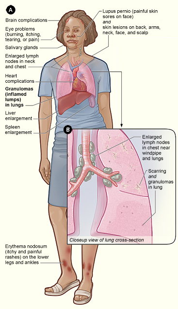
See here for a review of sarcoidosis, the "great mimicker".

Thursday, July 29, 2010

HOCM


Today we had a very information special guest morning report from the medical consults team. They discussed a woman with a new systolic ejection murmur, found to have hypertrophic obstructive cardiomyopathy on echo. They also discussed the complications of HOCM in pregnancy.

HOCM is a familial cardiomyopathy that results in marked asymmetric left ventricular hypertrophy (predominantly anterior ventricular septum) and diastolic dysfunction. In severe cases there is left ventricular outflow tract obstruction during systole, because the anterior (usually) leaflet of the mitral valve and septum actually meet. You may hear the term "systolic anterior motion" or SAM - this is what it is referring to.

Consequences of HOCM range broadly - from no symptoms whatsoever, to CHF and atrial fibrillation, to sudden cardiac death. Shortness of breath on exertion is the most common symptom. Predicting who will suffer serious consequences is difficult, but there are some generally agreed upon risk factors. These include:
  1. Family history of HCM with sudden cardiac death
  2. History of syncope or presyncope
  3. Massive LVH (septal wall thickness >30 mm)
  4. Previous (survived) sudden cardiac death
  5. Nonsustained ventricular tachyarrhythmias (NSVT)
  6. Abnormal blood pressure response to exercise
Physiologically we discussed how decreasing preload, increasing contractility, and decreasing afterload all worsen outflow obstruction.

Treatment also varies based on clinical presentation. Low risk asymptomatic patients do not require treatment. Betablockers and calcium channel blockers have a role in the symptomatic patient. Patients with CHF will likely require diuretics but this must be done cautiously as decreasing preload will worsen the outflow obstruction. Implantable cardioverter defibrillators can be used to prevent sudden cardiac death in a subset of high risk patients. Surgical treatment is generally reserved for patients who fail medical management and remain highly sympomatic.

See here for a review of management and risk stratification.

For those of you interested in preparticipation screening of athletes, here is an interesting article.

Have a good weekend and see you in a week!

Tuesday, July 27, 2010

Acute kidney Injury and HIV


Today we discussed an interesting patient with acute kidney injury in the setting of HIV, HCV, and DM II.

Acute kidney injury occurs more frequently in HIV+ patients than in HIV- individuals. This is distinct from the increased frequency of chronic kidney disease we sometimes see as the result of the HIV-associated glomerulonephropathies (ie HIV-associated nephropathy, IgA nephropathy).

The approach to renal failure remains the same: prerenal, renal, and postrenal. However, there are some specific causes that must be weighted more heavily or added to this list as a function of the patient being HIV+. For example:

Aside from the usual, prerenal causes must also include:
Pancreatitis (leading to 3rd spacing)
Cirrhosis/hepatorenal syndrome
Diarrheal illnesses

Renal causes must also include:
ATN
prolonged prerenal failure (not specific to HIV, but still the most common cause for ATN)
amphotericin B
aminoglycosides
pentamidine
ritonavir
cocaine

Acute tubulointerstitial nephritis
Several Abx, H2RB (ie ranitidine), NSAIDs

glomerular
HIVAN
IgA nephropathy
Immune-complex GN (ie cryoglobulinemia in the setting of HCV coinfection)

vascular
TTP, HUS
drug-induced
infection

crystal-associated nephropathy
sulfadiazine
indinivir
acyclovir

Aside from the usual, post-renal should also include:
nephrolithiasis (increased suspicion)
urethritis
retroperitoneal disease (ie lymphadenopathy)
infections (including fungal, TB-suspect in aseptic pyuria)

See here for a review that outlines the broad ddx.

Lastly, this study here looked at ARF in hospitalized HIV+ patients before and after the advent of HAART and found that HIV+ patients were more likely to have CKD, but also more likely to have ARF before and after HAART (adjusted OR 4.62 (4.3-4.95), and 2.82 (2.66-2.99), respectively. Age >65, DM, CKD, acute/chronic liver disease, and hepatitis co-infection were all statistically significant risk factors for development of ARF - interesting given that our patient had many of these. Interestingly, the incidence of acute renal failure was higher in the post-HAART era. Not surprisingly, in-hospital mortality was higher in HIV+ patients with ARF than in those without (OR 5.83 (5.11-6.65). Of course, it is difficult to say whether ARF is an independent predictor or more a marker of more severe illness.